What are the benefits of using AutoCAD in mammography?
CAD in mammography clearly increases the efficiency and confidence level of radiologists while searching for subtle microcalcification clusters and, when asked, most users will subjectively comment that they are less fatigued at the end of a CAD-supported reading session.
What is a CAD mammogram?
When reviewing your mammogram report or your invoice, you might come across the term “CAD”. CAD stands for Computer Assisted Detection, and it can be used to look at all sorts of different images, from x-rays to CTs. CAD uses specially designed software to analyze mammography images.
What are the benefits of CAD in cancer screening?
Most studies published to date suggest that there is a clear benefit to the use of CAD in terms of increased cancer detection rates, in particular with less experienced and/or low-volume reviewers. The actual demonstrated benefit increases from none to approximately 20% with most studies suggesting an increase between 2% and 10%.
What is the use of a computer aided detection mammogram?
CAD or computer aided detection is a computer-based technology that helps the radiologist in identifying suspicious areas while reading a digitalized mammogram. It was approved by the FDA in 1998. No randomized trials have been performed to assess its effect on breast cancer mortality.

What does CAD mean in a mammogram?
Advertisement. A large study suggests that using computers to help read older women's mammograms – called computer-aided detection (CAD) – means more invasive breast cancers are found earlier and more DCIS (ductal carcinoma in situ) is found.
How accurate is CAD on mammogram?
Diagnostic accuracy was not improved with CAD on any performance metric assessed. Sensitivity of mammography was 85.3% (95% confidence interval [CI]=83.6–86.9) with and 87.3% (95% CI=84.5–89.7) without CAD.
Is CAD required for mammography?
Must the use of CAD be dictated in the report? No, an order is not required for the use of CAD performed in conjunction with breast imaging procedures, such as mammography, MRI, and ultrasound.
Are 3D mammograms better for dense breasts?
A 3D mammogram offers advantages in detecting breast cancer in people with dense breast tissue because the 3D image allows doctors to see beyond areas of density. Breast tissue is composed of milk glands, milk ducts and supportive tissue (dense breast tissue) and fatty tissue.
Why does CAD fail in mammography?
In summary, we believe CAD failed because of insufficient process- ing power and supervised learning. Its widespread implementation unmasked the lack of its effectiveness. developed on a certain data set—for example, one image-view mammograms—are applied to different data sets [25].
Is CAD a 3D mammogram?
When paired with mammography, CAD helps radiologists identify any abnormal areas in breast tissue. To date, it's been used mainly with 2D imaging, but work is underway to extend its utility to 3D imaging, as well.
What does CAD mean in radiology?
Computer aided detectionSummary. Computer aided detection (CAD) is a clinically proven technology that increases the detection of breast cancer by assisting the radiologist in decreasing observational oversights (i.e. decreasing the false negative rate).
Is ultrasound better for dense breasts?
Ultrasound was slightly better at detecting cancers in dense breasts than 3-D mammography and both screening methods had similar false-positive rates. The study was published online on March 9, 2016 by the Journal of Clinical Oncology.
Why do I need a bilateral mammogram?
Bilateral mammography In addition to fat, breasts contain supportive tissue, blood vessels, ligaments, milk-processing glands and ducts. When these overlap on an x-ray, they can obscure tumors and other signs of breast cancer, leading to false negatives.
What type of mammogram is best for dense breasts?
Radiologists at RAYUS suggest that if you have dense breasts and fall into the “intermediate risk” category because of family history, you should consider 3D digital mammography (also called tomosynthesis). This imaging complements the standard 2D mammography and is performed at the same time.
Why do I need a breast ultrasound after a 3-D mammogram?
If you feel a lump in your breast, or one shows up on your mammogram, your provider may recommend an ultrasound. A breast ultrasound produces detailed images of breast tissue. It can reveal if the lump is a fluid-filled cyst (usually not cancerous) or a solid mass that needs more testing.
What is the best type of mammogram to get?
Breast health screenings that use digital mammograms have been proven to detect breast cancers better than conventional mammograms in three groups of women: those younger than 50, those with dense breasts and those who are pre-menopausal.
What does CAD mean in mammograms?
When reviewing your mammogram report or your invoice, you might come across the term “CAD”. CAD stands for Computer Assisted Detection, and it can be used to look at all sorts of different images, from x-rays to CTs. CAD uses specially designed software to analyze mammography images.
What is CAD in a radiologist?
CAD is designed to look for microcalcifications, areas of increased density and areas of asymmetry. In other words, it is looking for some of the same findings your radiologist looks for as signs of breast cancer.
What is a second look mammogram?
It is a “second look” at your mammogram to assist your radiologist in the task of finding subtle signs of breast cancer. Your radiologist interprets your mammogram fully, and then lets the CAD software analyze the images.
Does CAD improve mammography accuracy?
While there are studies showing increased accuracy of mammography using CAD, there are also studies showing it doesn’t alter accuracy significantly. It will never replace the trained and watchful eyes of your radiologist, but is a technical assistant.
What is CAD breast MR?
CAD for breast MR uses an entirely new class of temporal features. Its central feature is the automated analysis of time versus percent of enhancement curve using the market leader Confirma, which offers six different curve types highlighting the related parts of the entire lesion using six different colours. The curve type characteristics can be modified for each observer. The system of CADSciences is based on calculations of permeability and extracellular volume fraction and uses the calculations introduced by Toft. Thus, both technologies offer similar images but the underlining calculations differ significantly.
Why are CAD systems needed?
CAD systems are developed to assist the human reader in the detection of breast cancer. This development seems to be necessary due to financial and logistical problems associated with the double reading of radi-ologists.
Is CAD based analysis of breast MR images?
Till now , CAD-based analysis of dynamic breast MR images exclusively focus on dynamic data. It has been published recently, that morphologic features are of major importance as well, especially including T2w imaging. Thus, the currently available CAD systems do not yet replace the analysis of the entire examination, but shorten the evaluation of the contrast uptake only and induce a more reliable analysis. Depending on the presets, CAD systems are in part unable to highlight segmental enhancements, being the most relevant feature for non-invasive premalignant lesions. Diagnostic potential of currently available systems varies significantly. Whereas CADSciences technology is highly sensitive it seems to be significantly less specific compared to the currently available Confirma CAD solution.
Why is CAD important in mammography?
Because CAD is very sensitive and can detect very subtle abnormalities, it has proven to be particularly useful in mammography exams involving dense breast tissue, which is often present in pre-menopausal women. With the CAD technology, the radiologist still reviews all aspects of the mammogram and makes the final interpretation.
What is CAD in mammography?
Computer-aided detection (CAD) is a recent advance in the field of breast imaging and is designed to improve radiologists' ability to find even the smallest breast cancers at their earliest stages. CAD software uses sophisticated algorithms based on several thousand cases of breast cancer to identify suspicious areas on a mammogram ...
Can a radiologist diagnose a mammogram?
With the CAD technology, the radiologist still reviews all aspects of the mammogram and makes the final interpretation. CAD cannot diagnose. It simply serves as a highly valuable double check. As has always been the case, the most important thing is not the technology, but the person reading the results.
CAD in Screening Mammography
1 Both authors: Department of Radiology, Imaging Research, Ste. 4200, University of Pittsburgh, Magee-Womens Hospital, 300 Halket St., Pittsburgh, PA 15213-3180.
Recommended Articles
Pictorial Essay. MRI of Takayasu's Arteritis: Typical Appearances and Complications
Why do radiologists use CAD?
The goal was to ensure the radiologist did not overlook any visible structures when analyzing mammograms. CAD displayed loads of irrelevant data pinpointing all types of lesions, without distinguishing malignant and benign findings. Radiologists had to spend more time reviewing all these findings, which greatly slowed them down. Thus, a study revealed that a mammography interpretation assisted with CAD increased reading time by approximately 25 percent.
What is CAD software?
Computer-Aided Detection (CAD) is a diagnostic software that is based on traditional machine learning techniques. Machine learning employs algorithms to analyze information, learn from it and make informed decisions based on the knowledge gained. Compared to modern AI, traditional CAD uses more limited techniques that can only be trained on small datasets and is not capable of characterizing findings accurately. It is not capable of improving its performance independently by learning techniques by itself.
Does CAD help radiologists?
Studies evaluating CAD’s performance showed that it did not offer any significant benefits to radiologists. On the contrary, CAD negatively affected radiologists’ accuracy and productivity. Its use led to an increase in false-positive rate, which resulted in more needless patient recalls and biopsies, causing additional anxiety in patients.
Is CAD software on-premise?
Traditional CAD is known as on-premise software; it requires the installation of cumbersome equipment on each workstation and the on-sight presence of an engineer. Conversely, latest generations of AI systems are based on cloud technologies that require light on-site installation. These installations can be supported remotely, which provides for a decrease in integration expenses and time, and also provides the possibility of deployment during a pandemic situation such as COVID-19.
Case Summary
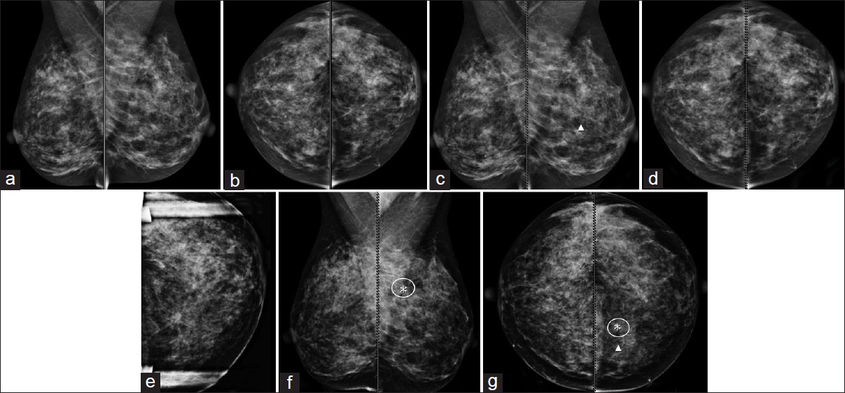
- A 74-year-old woman had a lump in the right breast that had three years earlier been identified as benign. Her daughter later became concerned that the lump had enlarged. Mammography accompanied by computer- aided detection (VuCOMP M-Vu® CAD), automated breast density (…
Imaging Findings
- Initially, craniocaudal (CC) and mediolateral oblique (MLO) views were done on both sides, along with a right true lateral view. These showed a rounded mass in the right retroareolar region corresponding to the palpable site. VuCOMP CAD (Figure 1) of the RCC identified the large mass, along with multiple areas of clustered, punctate, and pleomorphic microcalcifications. Calcificati…
Diagnosis
- Despite her initial reservations, the patient agreed to a biopsy after presentation of the mammography results. Pathology following ultrasound-guided core needle biopsy revealed grade 1 infiltrating ductal carcinoma in both breasts.
Discussion
- The value of computer-aided detection in breast cancer screening remains a topic of debate1, 2. A survey of radiologists by Siegel and Mezrich3 found that 87% of radiologists believe they would provide the same level of care without the use of CAD. Additionally, 62% of respondents claimed that they rarely or never alter their assessment after reviewing CAD results. To ensure CAD effic…
References
- Birdwell RL. The preponderance of evidence supports computer-aided detection for screening mammography. Radiology. 2009;253(1):9–16.
- Philpotts LE. Can computer-aided detection be detrimental to mammographic interpretation? Radiology. 2009;253(1):17–22.
- Elliot Siegel and Jonathan Mezrich, RSNA, 2013.